AI for early warning about liver cancer
 |
| AI can be utilised to detect the early signs of liver cancer |
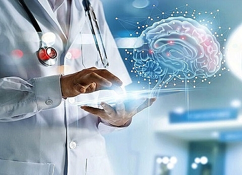
Associate Professor Vu Duy Hai, director of the Center for Biomedical Electronics of HUST, at the AI4VN conference taking place on August 15, said the centre is studying a system to support the diagnosis of liver cancer based on Big Data on ultrasound imaging and gene interpretation.
The research applying AI technology to diagnose liver cancer has been carried out since 2018 and will be fed ultrasound imaging data of 10,000 primary liver cancer patients. These images are collected from central and local hospitals of all ecological regions throughout the country. Of these, 200 patient records will be used to sequence genes to improve the accuracy of diagnosis.
After being transferred to the research centre, medical records from hospitals are standardised and labelled before storage. So far, the group has collected more than 6,000 ultrasound images.
After that, HUST will develop the tool to diagnose the early stages of primary liver cancer which can run on many devices like computers or smartphones. The application can be integrated into the ultrasound system of local medical facilities and hospitals.
"The application will be integrated into a software that can diagnose and screen early primary liver cancer with an accuracy of about 90 per cent," Hai said.
Liver cancer is one of the diseases with the highest mortality rates (40 per cent) in Vietnam. This research, if successful, will improve the chances of early diagnosis, giving many patients better chances at recovery.
However, doctor Dao Viet Hang from Hanoi Medical University Hospital expressed concern that ultrasound is not an accurate method to diagnose liver cancer, and that biopsy, tomography, or magnetic resonance all yield more reliable data. "Ultrasound images showing tumours cannot confirm that it is liver cancer. This application should only be used to screen for potential liver cancer and issue timely warning," Dr Hang said, suggesting that diagnosis and treatment should include a combination of health professionals and AI experts.
Dr Tran Quy Ha from the Dig Data research institute of Vingroup, argued that an image can be evaluated differently, depending on the doctor. Therefore, in the future, a combination of a council of doctors and the application of AI will provide the most accurate diagnosis. However, he said, AI must be applied throughout the entire process of medical imaging diagnostics.
"Another challenge is that the images collected must be annotated by the doctor's council and labelled. Every picture must be diagnosed by a doctor with a view to the cause and the appropriate treatment," he said.
The Big Data research institute is developing medical diagnostic imaging databases to help physicians have a firmer basis to make conclusions about medical conditions. At the same time, Vingroup also aims to decode the genes of 1,000 healthy people, finding genetic variations to build up a genetic database of Vietnamese people. As soon as these databases are completed, they will be shared free of charge with hospitals and communities to serve diagnosis and treatment.
Dr Tran Quoc Long from Hanoi National University said that the university is also collecting data to build AI applications to interpret echocardiography images. According to Long, many countries having such applications, but the technology is still too expensive to purchase, about VND3-5 billion ($130,434-217,391). Therefore, Hanoi National University is collecting echocardiography images from hospitals to build its own AI application for analysis and interpretation. The application can measure heart condition, ventricular structure, atria, and alert for myocardial infarction.
However, there are many difficulties, especially in image labelling because each medical record consists of about 14 videos, each containing 60-90 images. The doctor must study all the images to label and store in the database.
What the stars mean:
★ Poor ★ ★ Promising ★★★ Good ★★★★ Very good ★★★★★ Exceptional
 Tag:
Tag:
Related Contents
Latest News
More News
- Foreign leaders extend congratulations to Party General Secretary To Lam (January 25, 2026 | 10:01)
- 14th National Party Congress wraps up with success (January 25, 2026 | 09:49)
- Congratulations from VFF Central Committee's int’l partners to 14th National Party Congress (January 25, 2026 | 09:46)
- 14th Party Central Committee unanimously elects To Lam as General Secretary (January 23, 2026 | 16:22)
- Worldwide congratulations underscore confidence in Vietnam’s 14th Party Congress (January 23, 2026 | 09:02)
- Political parties, organisations, int’l friends send congratulations to 14th National Party Congress (January 22, 2026 | 09:33)
- Press release on second working day of 14th National Party Congress (January 22, 2026 | 09:19)
- 14th National Party Congress: Japanese media highlight Vietnam’s growth targets (January 21, 2026 | 09:46)
- 14th National Party Congress: Driving force for Vietnam to continue renewal, innovation, breakthroughs (January 21, 2026 | 09:42)
- Vietnam remains spiritual support for progressive forces: Colombian party leader (January 21, 2026 | 08:00)


 Mobile Version